The Radiograph Based Density Project has the ultimate goal to create a system that can be readily used in both private practice and research. In the first phases of testing, numerous private practitioners have stepped forward to participate in the project. As testing progresses, scientists from different disciplines are added to the research team. Below are brief introductions to the team members.
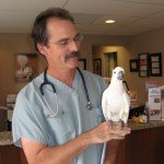
Dr Steve Barten
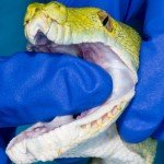
Toothless GTP with osteoporosis from Dr Steve Barten
Dr. Stephen Barten has always strived to contribute to the veterinary community. From his renowned clinical and wildlife photography to his involvement with the North American Veterinary Community, Barten has been prolific in the way he has shared his passion for reptiles. Barten is currently a veterinarian at Vernon Hills Animal Hospital in Mundelein, IL.
Barten is assisting his colleague Dr. Brad Waffa in the investigation of several rare cases of green tree pythons with osteoporosis and complete tooth loss. Bone density issues are common in reptiles such as lizards, turtles, and crocodilians. In these species, their problems are often related to improper diets or insufficient UVB light. Snakes, however, do not suffer from the same dietary deficiencies and most seem to thrive without UVB light sources.
“Most captive snakes are fed whole rodents, the bones of which provide adequate calcium and the livers of which provide adequate vitamin D, so bone density issues in snakes are very uncommon,” he said.
Barten has been involved with the North American Veterinary Community (LINK: http://navc.com/) for almost 30 years, serving as the reptile program chair, exotics coordinator, and now as the program coordinator for the annual conference.
“NAVC is the largest veterinary conference in the world with over 17,000 in attendance, including over 1,000 from more than 80 foreign countries,” he said.
Dr. José Biascoechea
Dr. José Biascoechea
Throughout his career, Dr. José Biascoechea has taken radiographs of hundreds of birds. Despite finding many anomalies in their bones and joints, quantifying the degree of the damage has been difficult because of variation between x-ray systems. Through the Radiograph Based Density Project, Biascoechea has been able to utilize specialized templates to improve our understanding of bone pathology.
“I have always wondered about the deficiencies that birds have when living in an indoor environment, not only from an enrichment point of view, but also from UVB deficiency and the overall health consequences that it leads to,” he said. “Scott contacted me to see if I would be able to help in his study, and without hesitation I agreed.”
Biascoechea is the founder of Exotic Vet Care in Mount Pleasant, SC. He is also the staff veterinarian for The Center for Birds of Prey and provides consulting and relief services for the South Carolina Aquarium and The Turtle Survival Center.
Dr Greg Burkett
Dr. Greg Burkett is the owner of The Bird Hospital: Avian Veterinary Services in Durham, NC. Currently, Burkett is in the process of purchasing a 3D scanner and printer so that he can develop prosthetics that are individualized for each bird he treats. He hopes that by 3D printing prosthetics, he can provide birds with stronger, lighter, and more natural options.
“The prosthetics are going to be true to form,” he said. “When I make prosthetic beaks by hand, they’re functional. But if I can 3D print them, I can get them almost to the cell level, and ultimately it will be more comfortable for the bird. For example, ducks have interlocking filtering mechanisms on their beaks, and I’ll be able to reproduce those with a 3D printer so that they can eat more naturally.”
Recently, Burkett treated a green cheek conure that presented initially with a fractured wing, but since then has broken the other wing as well as one of its legs, likely related to a hormone disorder. He said the Radiograph Based Density Study could help him visualize the rate of fracture healing, the quality of the healed bone, and whether other bones are likely to fracture soon.
“[This project] is giving veterinarians the ability to get more detail out of a radiographic film than we’re able to now by looking at the shades of grey more closely,” he said. “This may help with visualizing cancerous growths, osteoporosis, or numerous other bony lesions.”
Dr Angela Lennox

Dr Angela Lennox
Angela M. Lennox is a graduate of Purdue University School of Veterinary Medicine, and has practiced exclusively exotic animal medicine since 1991. She is the owner of the Avian and Exotic Animal Clinic of Indianapolis (www.exoticvetclinic.com). She is board certified through ABVP in both Avian and Exotic Companion Mammal medicine, and through ECZM in Small Mammal Medicine. She was awarded Exotic DVM of the Year at the International Conference on Exotics in 2005. Dr. Lennox is an adjunct professor at Purdue University Department of Clinical Sciences and teaches various exotic animal medicine topics to both veterinary and veterinary technician students. Dr. Lennox is past president of the Association of Exotic Mammal Veterinarians, editor of the Handbook of Rabbit and Rodent Dentistry, and co-author of Clinical Radiology of Exotic Companion Mammals. She is the AEMV section editor of the Journal of Exotic Pet Medicine. She resides near Indianapolis with her husband of more than 20 years and four daughters.
Dr Larry Nemetz
Dr Larry Nemetz
A pioneer in avian medicine and imaging, Larry Nemetz has envisioned the role of digital radiology in the avian world since 2005. Since that time, Nemetz has seen diagnostic imaging widely adopted by the veterinary field.
“In 2004 to 2006, [avian digital radiology] was feared, and now it is embraced but still not fully adopted in many practices,” Nemetz said. “The bad part for exotic practice is that the overall results of the images have not got better, which I expected, but instead have gotten worse because companies concentrate on the large [and] small animal field and not the small exotic realm.”
Since 1987, Nemetz has run his avian-only practice: The BIRD Clinic in Orange, California. In his practice, digital imaging has made a huge difference in the diagnosis and treatment of reproductive pathology and foreign body ingestion cases. Some of Nemetz’s radiographic images of grey parrots have been utilized as part of the Radiograph Bone Density Project.
“Anatomy is fluid, not fixed, so it can change and mold dependent on pathology,” Nemetz said. “It is always cool to see something you have never seen before in a given species or in general.”
Beyond digital imaging, Nemetz has interests in microsurgery and reproductive disease. In 2015, Nemetz traveled and lectured on avian medicine in Spain, France and Texas, and consulted in avian surgery in the United Kingdom.
Dr Drury Reavill
Dr Drury Reavill
Dr. Drury Reavill is the owner of Zoo/Exotic Pathology Service in Carmichael, CA, a specialty lab focusing on the pathology of non-domestic species. She has been helping create awareness for the Grey Parrot Anatomy Project and Radiograph Based Density Project within the veterinary community, and has been working on providing access to avian digital images for an upcoming exotic animal pathology book.
“The Radiograph Based Density Project has come into play with our recent work on evaluating green tree pythons for loss of their teeth and the fact that they can no longer replace the teeth,” she said.
Dr. Reavill has over 30 years of experience in avian and exotic clinical practice, laboratory diagnostics, and pathology. She completed a residency in anatomic pathology, has board certification with the American college of Veterinary Pathology (ACVP), and is a charter diplomate of the American Board of Veterinary Practitioners (ABVP) with avian specialty status. She has recertified in ABVP avian practice twice and has also obtained ABVP diplomate status in Reptile & Amphibian Practice.
Reavill has authored or co-authored over 150 refereed and conference papers, 20 book chapters, two books, and a CD, each covering a wide range of exotic animal disease conditions.
Dr Brian Speer
Dr Brian Speer
A pioneer of modern avian medicine, Dr. Brian Speer has worked with both the Grey Parrot Anatomy Project and the Radiograph Based Density Study since their inception. His research focuses on enhancing the understanding of avian skull anatomy, muscular anatomy, kinesiology, and their applications to clinical practice.
“In reality, it was this original interest that sparked Scott (Echols) and my exploration into the types of CT imagery that was available, and got us started on the road to where the project has headed today,” Speer said.
Speer’s findings have allowed the researchers to balance prokinetic function, keratin form, and wear, with myology and functional kinesiology. This has improved his approach to complicated clinical issues of the beak, such as treating lateral beak deviations in large macaw species, prognathism in cockatoos, and keratin overgrowth issues.
“Probably the largest change in our thought processes that has come from our research paired with dissections is the ability to balance prokinetic function, keratin form and wear, with myology and functional kinesiology as we work to correct the problems that we see,” he said. “The fascinatingly complex nature of prokinetic and coupled kinetic function has actually become more dynamically interactive and clear.”
As part of the density project, Speer has also been taking HD CT images of patients and specimens at his clinic, The Medical Center for Birds, in Oakley, California.
“The nature of my work is predominantly clinical practice, although I teach regularly at my alma mater, the [University of California Davis], and I am an author,” Speer said. “That of course, is balanced with trying to be a good dad, and husband to my best friend in life, Denise.”
Speer’s most recent publication is Current Veterinary Therapy in Avian Medicine and Surgery.