Skeletal:
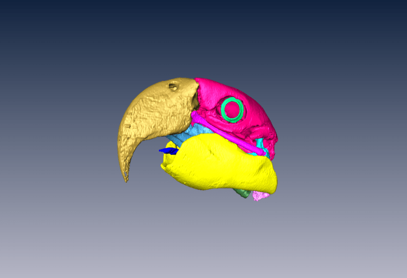
Digital segmentation of the bones of the head of a large macaw (Ara sp)
As a part of ‘proof of concept’ we needed to show that the individual bones could be digitally segmented from the CT data sets. The bones were digitally and painstakingly outlined one frame at a time to determine all of the margins. The segmented bones then become their own file (rather than attached together as one large file) and can be viewed in 3-D to study all of the detailed features. Here the individual bones are color coded and displayed in situ (in normal position). While time consuming, this technique allows us to digitally build a skeletal framework on which we can later add muscles, blood vessels, etc. Created from 100µm digital slices.
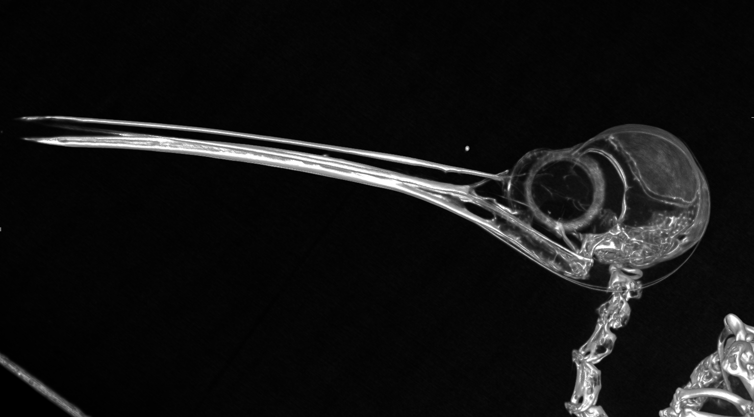
Digital reconstruction and video rotation of the grey parrot (Psittacus erithacus erithacus) head and neck. The rotation comes from one of our earlier attempts at digital reconstruction of micro CT images. The double line across the back of the skull is an artifact and later models show continuous bones. 100µm digital slices.
Skull and beak of a hummingbird (Archilochus sp.)
The bird was brought in dead to a local hospital. Notice the large eye to skull area and hyoid apparatus that extends from the base of the tongue to the dorsal rostral aspect of the skull. The tongue literally wraps around the head anchoring at the top front portion of the skull. This allows the hummingbird to extend the tongue far beyond the tip of the beak and into a flower. When retracted, the base of the tongue literally wraps around the back of the skull. 40µm digital slices.
Small birds were used to develop settings on the micro-CT. The budgie was perfused with BriteVu® and then scanned on a micro CT scanner. The budgie was scanned at 29µm digital slice thickness providing incredible detail.
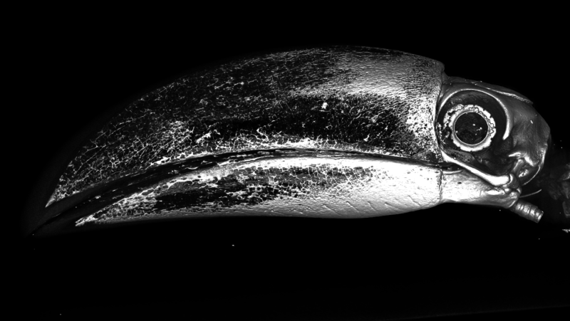
Lateral view of a keel-billed toucan (Ramphastos sulfuratus) head
This toucan was deceased and was used as a model to help another closely related toucan with a beak fracture. Notice the trabecular pattern on the upper and lower mandibles giving great strength and minimal weight. The tips of the upper and lower mandibles are covered with thick keratin. Based on the techniques we have developed across a spectrum of birds, we are able to perform micro and regular CT on a large variety of bird species with amazing results. 100µm digital slice.
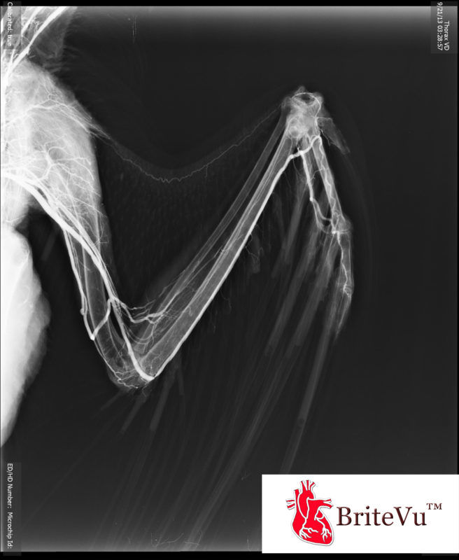
Golden eagle (Aquila chrysaetos) arteriovenogram
This eagle was euthanized (terminal condition) and entered into the study for arteriovenogram development. Out of similarly donated birds, a new technique and product (BriteVu®, research contrast media) to highlight the vascular system has been developed by Dr. Scott Echols. BriteVu has been used in this Project and many other studies. This image is a flat radiograph (‘X-ray’) that clearly shows the blood supply to and from the wing. Notice the blood supply along the prepatagial tendon (front of the wing web), distal phalynx of the minor digit (last bone at the tip of the wing) and the individual emerging (blood) feathers. Standard digital radiograph.
.")
The foot of a pigeon (Columba livia).
The pigeon was humanely euthanized (feeder bird) and donated to the project. Using a newer contrast agent (BriteVu®, research contrast media) and injection technique, even small vessels (to the ends of the digits) are readily visualized. Notice how the blood supply dramatically tapers off at the base of the claw (at the end of the digits). This and other similar images help us understand where the blood supply (and presumably the nerve supply) starts and stops at the extremities of birds. Much work still needs to be done but helps researchers understand the welfare issues associated with beak and nail trimming and just how much is ‘too much’. Also, this highlights that main aterial-venous blood ends at the base of the nail. More distal (towards the tip of the nail) blood is likely capillary and too small to see on this view. Using nail trims to collect blood for diagnostic sampling has long been suspected to represent capillary blood (which has some different lab values compared to blood from non-capillary sources). Images from several birds species support that nail sampling is indeed capillary blood and caution should be used in its clinical interpretation. 100µm digital slice.
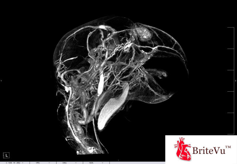
Sagittal (digital) section through a grey parrot (Psittacus erithacus erithacus) head post-arteriovenogram.
Sagital section of the head and neck of a grey parrot (Psittacus erithacus erithacus) post ateriovenogram (BriteVu®, research contrast media). This bird was euthanized (terminal condition) and donated to the project. This single bird has provided an amazing amount of data most of which has been through exquisite arteriovenogram images. Dr Echols developed a new product and technique that was used to create an arteriovenous ‘cast’ that was then easily viewed on micro-CT. From the head alone we have learned about several new features: 1.) A large blood supply going to the nares (nostrils) 2. Understanding of the blood supply to the upper and lower mandible 3. What appears to be a venous plexus at the base of the brain. Again, much research is needed here but it opens up a better understanding of surgical approaches to the beak that best preserve blood supply, how the parrot brain regulates temperature and how nutrient exchange occurs in parrot brains without direct access to a large blood vessel. 100µm digital slice.
Muscles:
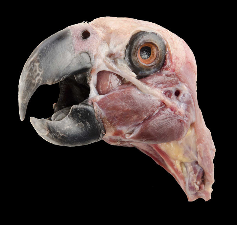
Grey parrot (Psittacus erithacus erithacus) superficial dissection of the head.
Superficial dissection of the grey parrot (Psittacus erithacus erithacus) head. This bird was euthanized (terminal condition) and donated to the project. Initial detailed dissections of the grey parrot have already revealed significant anatomic variations from other published bird species. Even within parrot species, we are finding significant variation in muscle anatomy. Despite the variations we have encountered, there is enough similarity for properly naming the muscles. Detailed dissections such as the one viewed here will be used to determine origin and insertion and proper naming based on currently reported anatomy in the Nomina Anatomica Avium (Handbook of Avian Anatomy). Picture courtesy of Mark Nielsen.
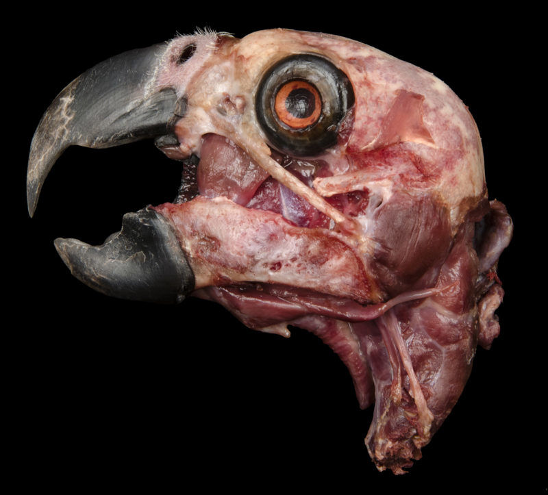
Deeper dissection of the same grey parrot above.
Mid-level to deep dissection of the grey parrot (Psittacus erithacus erithacus) head. This bird was euthanized (terminal condition) and donated to the project. The exposed mandible and jugal bone are readily visible. The Pseudomasseter muscle is triangular with the base located on the boney orbit (eye socket) and the point on the medial aspect of the mandible (inside the jaw bone). This muscle is thick, strong and elevates the lower mandible. This muscle alone has been highly variable among parrot species. Knowledge of this and other muscles not only helps with anatomy studies but is used clinically as a landmark when performing surgery of the suborbital chamber of the infraorbital sinus (air space immediately beneath the eye). This happens to be a common area of infections in parrots and other birds. Picture courtesy of Mark Nielsen.
This bird was euthanized (terminal condition) and donated to the project. Our initial attempts to visualize muscles using advanced imaging techniques have not proven successful. In other animals where MRI or CT have been used to image the muscles, those species tend to deposit fat in between muscle layers improving visualization. Parrot species tend to lay fat around internal organs and under the skin (when overweight) and do not offer visualized separation of muscle with the current technology. As a result, we are using hand performed dissection as the most reliable means to identify muscles. As a side note, this bird has very poor bone density and can best be seen in the skull as irregular ‘holes’ in the bone. These ‘holes’ represent areas where the bone tissue is less dense than the muscle that can still be seen surrounding the bones of the main body. 100µm digital slices.
Internal Organs:
This cockatoo was euthanized (terminal condition) and the body donated to the project. The blood supply to the internal organs was filled with an older contrast agent and technique. Regardless, we were able to see the blood supply within the heart at great detail. The atria are filled with contrast agent while the ventricles are empty (bottom of heart). The top of the image shows the major vessels coming off the heart base. Images such as this have helped us better understand how to improve the contrast technique and visualize the vasculature to various internal organs of birds at detail never before seen. 100µm digital slices.
The pigeon was humanely euthanized (feeder bird) and donated to the project. The vasculature of the lungs is highlighted with contrast agent (BriteVu®, research contrast media) creating a tree like appearance. The lungs of birds are fixed and therefore expand very little (much unlike with mammals). The intense vascular supply is needed to help exchange air as it flows through the lungs rather than as the lung expands (mammals). This study gives us a clear view as to how blood courses through the lungs of at least one species of bird. It also gives us the outline of the lungs. 100µm digital slices.
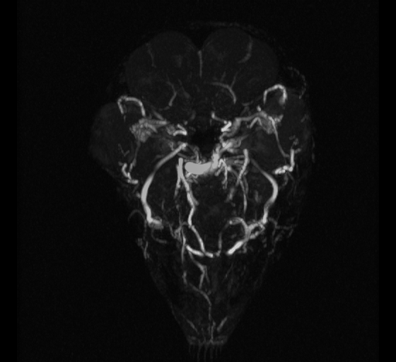 Time of flight is a relatively new MRI (Magnetic Resonance Imaging) technique that allows researchers to directly visualize the blood supply and flow in living animals with no contrast agents. Essentially this is a non-invasive means to look at blood vessels and study blood flow. We are currently developing this technique to non-invasively evaluate normal and abnormal vascular structures. The video you see shows the head (beak tip or rostral to the left and caudal or back of head to the right) of a blue and gold macaw (Ara ararauna) with a severe sinus infection.
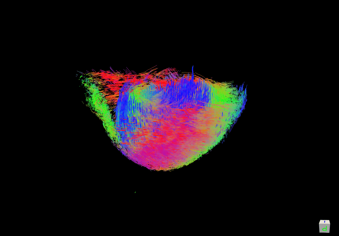 Cardiac myocyte mapping is another new technique that allows researchers to ‘see’ each individual muscle cell within an organ- in this case the heart. The organ is imaged using MRI and must be harvested from a deceased animal at this time. The results (see the video) show individual muscle cells (streaks) and groups of cells in a region (designated by common colors). The images give a 3-dimensional view of how the muscle cells are oriented in space. In normal tissues, this allows researchers to ‘see’ how muscle cells contract and in what direction giving anatomical and physiological information as to the muscle as a whole. In abnormal tissue it shows where the muscle contraction process has gone wrong and helps us understand the disease better. The video shows the lower (distal or apex) half of the normal heart of a Moluccan cockatoo (Cacatua moluccensis).