The Comprehensive Anatomy Research Project serves not only to discover ‘anatomy’, it also was created to develop diagnostic and treatment technologies that can be used here and now on living animals (including humans). Team members come from many walks of life and scientific disciplines. Each researcher brings a unique set of skills and, in some cases, a student force to help tackle problems encountered through the research process.
Dr. Scott Echols
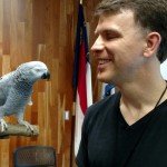
Dr. Scott Echols- Principle Researcher, Project Coordinator, Co-Author
Dr. Echols is a board certified avian specialists with over 25 years of experience in practice, teaching and research. His main interests are avian surgery, nutrition, kidney disease and improving the behavioral health of captive species. Dr. Echols is the co-founder of Mobile Avian Surgical Services, founder of Avian Studios and Scarlet Imaging, a frequent author and lecturer and has created educational DVD’s and other videos that have been viewed worldwide.
Dr. Justin Adams
Dr. Justin Adams, Monash University. Melbourne, Australia
Dr. Adams has an interdisciplinary background in human and comparative anatomy and mammal palaeobiology while leading field and lab research teams over the past 17 years. He has published over
50 peer-reviewed papers and led 16 competitive internal and external grants since 2002 across research in palaeontology, 3D printing, and comparative anatomy. He is a world recognised expert in the anatomy and evolution of African mammals, including leading faunal analysis at several fossil human sites in South Africa and in the applications of imaging and 3D methods in the discipline. His research and publication background in applications of imaging (CT, μCT, MRI, surface scanning) analysis and interpretation has been central to the publications on different mammal species with a more recent focus on Australian marsupials.
Brent Adrian
Senior Research Specialist, Department of Anatomy, Midwestern University
Brent Adrian is a Senior Research Specialist in the Department of Anatomy at Midwestern University. He earned a Masters of Fine Arts in painting and drawing from Arizona State University in 2006 and was hired by the University of Arizona College of Medicine- Phoenix as a medical illustrator. His work as a researcher focuses on the functional anatomy of rare and threatened mammals, and his illustrations have appeared in numerous scholarly publications. In 2012, he joined the research staff of the Department of Anatomy at Midwestern University, where his work in describing and illustrating the anatomy of rare animals continues. He also studies fossils and has authored articles on turtles from the Cretaceous of Texas and Eocene of Utah, as well as Miocene carnivores of Africa. As an illustrator, he generates many of the original scientific illustrations in the ARCIVES publications.
Scott Birch

Scott Birch, 3D Art Design GPAP
Scott Birch has been focused on the technique of translating CT/MRI/PET data into interactive 3D models or visualizations for over two years, and his first concentration was on veterinary cardiology.
“In 2014, Dr. Ashley Saunders at Texas A&M had a heart murmur project that required visuals of specific congenital heart defects, and we were stumped,” he said. “I remembered a not-too-successful technique I had attempted in 2008 at Cornell University to visualize anatomy for regional nerve blocks, so I tried again with an updated toolset. Through many months of trial-and-error, I finally honed in my technique and was able to build 3D models successfully, visualize complex anatomy in an interactive environment, and even print replications of patient-specific anatomy.”
In this pursuit of knowledge, Birch needed extremely high resolution data sets to unlock the potential of his technique, and thus became involved with the Grey Parrot Anatomy Project.
“Dr. Echols has used his new contrast agent, Brite-Vu®, to perfuse a parrot and then CT scanned at 100 micron resolution, which was the highest resolution scan I had seen so far,” Birch said. “He shared with me over 17 gigabytes of data—an incredible puzzle for me to re-build as a 3D model. It took me months to segment the tissue, isolate the structures within, and add color and material to the 3D models.”
Once Birch has the 3D models, he can rotate them to any viewpoint, change transparency or translucency, add clipping planes, or otherwise modify them to yield the final render.
“These images are just as much art as science, and my goal is to make them as beautiful as possible while also showing the areas of interest to scientists and educators,” Birch said. “I am not a scientist, researcher, or bio-engineer, but rather an artist who is borrowing techniques from all of these different disciplines to produce my work.”
Birch hopes to 3D print the parrot’s vascular structure at 25x real size, or over 2 meters from head to claw if possible. He said the models can also be shared with other educators and researchers across the globe.
“One bird could be shared across the entire globe, essentially living forever,” he said. “I think that’s pretty cool.”
Jason Bourke
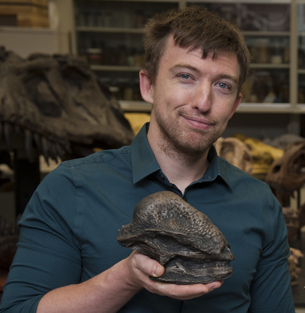
Jason Bourke, Arkansas State University
Jason Bourke is an Assistant Professor of Anatomy in the Basic Sciences department at NYITCOM at Arkansas State University. Bourke studies the evolution of thermoregulatory structures in amniotes, with a particular focus on the paleophysiology of dinosaurs. To reconstruct lost soft tissues, he looks at the anatomy of modern relatives of dinosaurs (birds, crocodylians, and lizards). Shared anatomy is inferred to have been present in the extinct animals. To test aspects of thermoregulation, Bourke uses computational fluid dynamics to simulate the movement of air, blood, and water. This allows him to follow heat-flow pathways and test the efficiency of different anatomical structures at moving heat to and away from parts of the body.
The nose of dinosaurs has proven particularly good at handling heat, with large dinosaurs from multiple distantly related groups evolving equally large noses. “This indicates that nose elaboration may be inextricably linked to body size evolution” Bourke said. To further understand this pattern, Bourke is looking at monitor lizards (Varanus), which have the largest body size range of any animal today (smallest to largest differ by over 4,000x in mass). “Such a vast range in body size is bound to create different thermal problems at the extremes of that range.” By studying the nose shapes of different monitor lizards across their size range and environments, Bourke hopes to unravel the underlying mechanisms that lead to nasal elaboration and understand just how coupled this anatomy is to body size.
For more details on Dr. Bourke, you can visit his website: https://www.bourkelab.com.
Dr. Margaret Hall
Dr. Hall earned her Ph.D. in Anatomical Sciences from Stony Brook University, and is a member of the Anatomy Department at Midwestern University where she teaches both veterinary and human anatomy. Dr. Hall is a comparative anatomist with research exploring important anatomical features of vertebrates. One major focus of her research is the evolution of vertebrate visual system morphology, including eye shape, extraocular muscles, and retinal morphology. A second focus area is the evolutionary history of the mammalian body wall – including humans – especially as muscular morphology extends from the thorax and abdomen and relates to perineal structures that result from cloacal septation, an important adaptation in mammals.
Dr. Casey Holliday
Dr. Casey Holliday, associate professor of anatomy at the University of Missouri School of Medicine, combines techniques from different fields to understand the biomechanics and evolution of the skulls of birds and crocodilians.
Using iodine contrast-enhanced micro CT imaging, histology, and biomechanics, the Holliday lab has created 3D models of parrot heads to study cranial kinesis.
“Cranial kinesis is the phenomenon that parrots and many birds, snakes and some other animals have in which they can move joints in their heads other than just the jaw joint,” Holliday said. “A parrot can elevate its beak across the hinge joint its face, so it can husk fruits and nuts, or use its bill to climb around on trees. There are all these extra mobile joints and muscles that allow them to do this behavior which I find fascinating. Parrots are pretty phenomenal example of kinesis.”
By creating a 3D model, the researchers can shed light on how parrots are able to generate such high bite forces and move their bills dexterously. They are also able to see how different muscles load force at different points in feeding. Some of this data will be used as part of the Grey Parrot Anatomy Project.
Ian Cost, a PhD student at the Holliday lab, is currently studying adaptations to bone and cartilage among different bird species to understand how some birds are able to generate high forces in their heads without breaking bones. For example, ducks have flexible bones in their jaws that can bend up to 30 degrees without breaking. Parrots, on the other hand, need to have extremely strong bones in order to crack open nuts with ease.
Recently, the lab has also been able to compare CT images of the Kea and Kakapo, both parrots from New Zealand.
“Grossly speaking, you can see just from their head shape that they can do really really different things with their heads,” Holliday said. “They are more closely related to each other than they are to other parrots, so the argument can be made that their anatomy diverges so much so that they can occupy different ecological niches.”
While most of the lab’s work is in the realm of basic sciences, Holliday said eventually the research could have clinical applications for treating disease processes like arthritis, fractures, or beak malocclusions.
The Holliday lab’s research was recently featured on the cover of the Journal of Anatomy.
Dr. Dominique Homberger
Dr. Dominique Homberger is a comparative anatomist and Andrew Clinton Pereboom Honors Professor at Louisiana State University. She is currently in the early stages of analyzing the anatomical base of palatine luxation in parrots.
“My research on the palatine luxation research is a collaborative project with Scott Echols and Julie Hebert, who collected the first observations of this problem,” she said. “I plan to look into the anatomical reasons for this malfunction.”
According to her website, Homberger’s research projects are tied to a “fundamental interest in the reconstruction of macroevolutionary changes as a result of individual variation and natural selection by synthesizing functional-morphological and behavioral-ecological data of extant organisms with paleoclimatological and geological data.”
Beyond her palatine luxation research, Homberger has been working on a long-term study of feeding and drinking behavior, functional morphology, ecology, and evolutionary history of the Psittaciformes.
Jackie Houser

Jackie Houser, Oregon State University
Jackie Houser is a member of the DVM class of 2018 at Oregon State University. Houser is currently helping Dr. Sarah Nemanic with a interactive online project designed to teach normal radiographic anatomy of birds, dogs, cats, and horses.
“The program currently includes dog, cat, and horse radiographic anatomy, but I have an interest in avian medicine, so I joined with the intention of adding a few avian species to the program to help the bird lovers like me out there,” Houser said. “It’s designed to be interactive to help create an association between the structure on an image and the name of the structure, in order to help facilitate learning normal radiographic anatomy.”
Houser and Nemanic currently have normal radiographs from a red-tailed hawk, domestic duck, and chicken, but are hoping to expand the project by gathering more images of pet bird species.
“In order to be able to recognize something as truly being abnormal on a radiograph, you first need to know what is normal,” she said. “Since veterinary students don’t receive as much exposure to avian anatomy as we do other species, I thought it was important to try and expand the resources and tools available for learning normal avian radiographic anatomy.”
Houser has two dogs, a cat, and a Chinese water dragon at home. She enjoys reading, hiking, playing video games, and occasionally brewing beer. She hopes to work with pet birds and exotics once she graduates, as well as volunteer with local wildlife centers.
“I first became more interested in birds during [my undergraduate degree] at University of Oregon when I volunteered at the Cascades Raptor Center in Eugene,” she said. “For the past two years I have been volunteering at Chintimini Wildlife Center in Corvallis. I think birds are such fascinating creatures, and I would like to stay involved with wildlife conservation efforts.”
Dr. Jeremy Klingler
Dr. Jeremy Klingler, Assistant Professor, Biology, Southwestern Oklahoma State University
Dr. Klingler is an assistant professor of biology at Southwestern Oklahoma State University who specializes in vertebrate paleontology and more specifically, archosaur paleobiology. Klingler is highly interested in muscle anatomy and physiology and in reconstructing what dinosaur muscles were like and how they functioned. His research has recently focused mostly on the evolution of the neck and underlying related neck structures (e.g., the trachea and esophagus). In addition, he is very interested in a group of muscles in birds called dermo-osseous muscles. These muscles have received little attention but are likely to provide an interesting evolutionary story for the integumentary system of non-avian dinosaurs and birds. Klingler has been helping to dissect and describe the muscular system of the grey parrot.
Dr. Andrew H. Lee
Andrew H. Lee, Associate Professor of Anatomy, Midwestern University
I am an evolutionary morphologist with expertise in bone histology, biomechanics, and growth. This expertise gives me a unique perspective that integrates form, function, and time. With this perspective, I seek to understand why skeletons have certain microscopic features.
By using the annual growth lines preserved in dinosaur bones to estimate growth rates, my work shows that large dinosaurs matured about five times faster than living reptiles scaled to comparable size. Even small dinosaurs grew about 40% faster than living reptiles. A potential ecological advantage of rapid growth is the increase in lifetime reproductive success. These findings clarify how dinosaurs were successful for 150 million years as well as the precedent for rapid growth in their descendants, birds.
More is known about bone histology in extinct species than in living ones. Therefore, my current research focuses on living species. I am currently testing whether bone with circumferential vascular canals (laminar bone) is an adaptation to flight. Laminar bone is thought to resist twisting loads and is prevalent in some of the wing bones of birds. But it is absent in bats. The absence of laminar bone in bats is best explained by relatively slow growth compared to birds. This work suggests that growth is an important constraint on wing bone histology. To test this further, my students use a variety of tools including traditional histology, microCT, fluorescent microscopy, bone strain analysis, and principal component beta regression to understand how flight adaptations develop in growing birds. Recently, we reported that bone with longitudinal canals increases in the limb bones of growing pigeons, suggesting a novel adaptation. This finding coupled with future work will clarify how avian skeletons are adapted to flight and inform bio-inspired design.
https://www.midwestern.edu/academics/our-faculty/andrew-h-lee-phd.xml
Ashley Mooney
Ashley Mooney, writer for GPAP
Ashley Mooney is a veterinary student at the University of Melbourne and an aspiring avian specialist. She has been assisting the Grey Parrot Anatomy Project by liaising with researchers and writing promotional briefs for the Project’s website.
While birds are her primary passion, she has sought experiences with primates through the Jane Goodall Research Institute, where she did research on chimpanzee behavior. She has also worked as a science writer since 2010, first as a writer and editor for Duke University’s student newspaper, and now as a freelance editor.
Mooney is grateful for the opportunity to work with so many esteemed veterinarians and researchers through the Project, and is excited to contribute more to the field once she graduates.
Dr. Sarah Nemanic

Dr. Sarah Nemanic
Dr. Sarah Nemanic is an assistant professor of radiology at Oregon State University. Prior to attending veterinary school, Nemanic earned a master’s and Ph.D. in neuroscience in the field of learning and memory at the University of Texas, Houston Health Science Center. She has been a board-certified radiologist since 2011.
Nemanic is currently working on an interactive online resource for teaching normal radiographic anatomy to veterinary students. The project initially focused on dogs, cats, and horses, but has since expanded to include avian species. Veterinary student Jackie Houser and computer scientist Matt Viehdorfer are both assisting with the project.
“Creating the normal radiographic anatomy software application is a fusion of my interests in learning and memory and veterinary radiology,” she said. “We did an experiment testing the efficacy of this program on the dog radiographic anatomy and found that it was highly effective. This will be in an upcoming issue of the Journal of Veterinary Medical Education.”
Nemanic’s other research interests include methods of diagnosing orthopedic diseases in dogs and staging of canine and feline oncology patients.
Professor Mark Nielsen

Mark Nielsen- Anatomy Coordinator and Chief Anatomist, Co-Author
Mark Nielsen is an anatomy professor and frequent lecturer and author. As one of the most frequently published authors in human anatomy textbooks and digital applications, Mark is a highly sought after lecturer, writer, reviewer and researcher. Mark’s publications include animal anatomy and he has extensive experience in detailed dissection and comparative studies.
Dr. Emma Schachner

Dr. Emma Schachner- contributor, grey parrot respiratory system
Emma Schachner, an Assistant Professor in the Department of Cell Biology and Anatomy at Louisiana State University Health Sciences Center, has been collaborating with the Grey Parrot Anatomy Project since her days as a post-doctoral researcher.
Schachner’s primary focus is on the evolution of reptile and bird lungs. As a paleontologist and evolutionary anatomist by training, Schachner looks at modern relatives of dinosaurs to reconstruct their respiratory anatomy. With over 150 scans of several species, Schachner is working on qualitative descriptions of the air sacs, lungs, and other branches of their respiratory tracts. When working with engineers, Schachner is even able to visualize airflow patterns or transform her CT data into 3D-printed models.
“I’m interested in form and function, like why is something shaped the way it is and why it evolved to be that way,” Schachner said. “Every bird that I do a model of provides an exciting surprise to see how it’s going to turn out.”
Although Schachner’s research is broad in its nature, she said she prioritizes her studies on the grey parrot. Because of the Grey Parrot Anatomy Project, Schachner is able to utilize larger sample sizes (N = 9 birds) to overcome the challenges presented by individual variation, and create a more comprehensive view of the species’ respiratory anatomy. Additionally, the quality and power of the micro CT imaging allows her to build even more accurate high resolution models.
Eventually, Schachner hopes to scan a kiwi bird, which according to a publication from the 19th century, does not have an abdominal air sac like other birds. She also said she would love to image a hoatzin, a pheasant-like bird from South America. Hoatzin juveniles possess claws on their thumb and first finger, allowing them to climb trees. Schachner said that some of the genetic mechanisms that control limb development are linked to lung development, and she is curious as to how much the hoatzin respiratory tract differs from other birds.
For more details on Dr. Schachner, go to her website www.theropoda.com.
Dr. Nico Schoemaker and Dr. Yvonne van Zeeland
Dr. Yvonne van Zeeland and Dr. Nico Schoemaker from Utrecht University are working to unravel the mysteries of the neuroanatomy and neurophysiology of the African grey parrot.
Although a basic understanding of the avian brain has been established, there currently is relatively little information on brain pathology and functional linking of brain regions in birds. By using micro-MRI and Diffusion Tensor Imaging (DTI), Yvonne and Nico hope to elucidate neural pathways between different brain regions.
“This research will help to gain further detailed knowledge of the normal avian brain, following which comparison of healthy with abnormal brains may take place to help gain a better understanding of brain diseases in birds, and how these may be dealt with,” she said.
Eventually, Yvonne and Nico also hope to apply their findings to prevent and treat behavior issues such as feather damaging. This behavior problem is particularly common in grey parrots and cockatoos, with a reported prevalence of up to 40 percent.
“We are hypothesizing that this type of abnormal repetitive behavior is similar to compulsive/impulsive behaviors where there is a lack of inhibition from the centers that regulate and control the initiation of behaviors,” she said.
As a consequence of this lack of inhibition, birds may not be able to control urges to damage feathers, which may particularly be true in chronic cases. DTI may allow the researchers to visualize functional differences in the brains of birds that are expressing these behaviors versus healthy individuals and also allow comparison with those that exhibit stereotypic behaviors, which they hypothesize to be controlled by different brain regions.
Beyond their research on neuroanatomy, Nico and Yvonne are also studying the influence of the parrot’s personality and living environment on the likelihood of a bird developing feather damaging behavior. As part of this project, an online survey has been prepared for owners to fill out information regarding their bird, its behavior and living environment.
“With the help of the information derived from this and other studies, we hope to gain insight in how to tailor environmental and social enrichment to an individual parrot’s behavioral needs, thereby hopefully also preventing behavior problems such as feather damaging behavior,” she said.
More information about the studies conducted in Utrecht is available on the Utrecht University webpage.
Dr. Heather F. Smith
Heather Smith is a Professor of Anatomy and the Director of Anatomical Laboratories at Midwestern University and one of the coordinators of the Arizona Research Collection for Integrative Vertebrate Education and Study (ARCIVES). She is an anatomist and evolutionary biologist who studies evolution and functional morphology in mammals and turtles. Her recent work on extant carnivorans has focused on functional morphology of locomotion and anatomical adaptations of the limbs. Her paleontological work focuses on the evolution of turtle communities in the Western Interior of North America, in particular from the Late Cretaceous through the Eocene. Her research on primates has centered around the relationship between cranial morphology and phylogeny, environment, and diet. Dr. Smith is the Principal Investigator on the snow leopard and tortoise projects, conducted in collaboration with other researchers at MWU and Dr. Scott Echols.
https://www.midwestern.edu//academics/our-faculty/heather-smith-phd.xml
Dr. Brian Speer
Dr. Brian Speer
A pioneer of modern avian medicine, Dr. Brian Speer has worked with both the Grey Parrot Anatomy Project and the Radiograph Based Density Study since their inception. His research focuses on enhancing the understanding of avian skull anatomy, muscular anatomy, kinesiology, and their applications to clinical practice.
“In reality, it was this original interest that sparked Scott (Echols) and my exploration into the types of CT imagery that was available, and got us started on the road to where the project has headed today,” Speer said.
Speer’s findings have allowed the researchers to balance prokinetic function, keratin form, and wear, with myology and functional kinesiology. This has improved his approach to complicated clinical issues of the beak, such as treating lateral beak deviations in large macaw species, prognathism in cockatoos, and keratin overgrowth issues.
“Probably the largest change in our thought processes that has come from our research paired with dissections is the ability to balance prokinetic function, keratin form and wear, with myology and functional kinesiology as we work to correct the problems that we see,” he said. “The fascinatingly complex nature of prokinetic and coupled kinetic function has actually become more dynamically interactive and clear.”
As part of the density project, Speer has also been taking HD CT images of patients and specimens at his clinic, The Medical Center for Birds, in Oakley, California.
“The nature of my work is predominantly clinical practice, although I teach regularly at my alma mater, the [University of California Davis], and I am an author,” Speer said. “That of course, is balanced with trying to be a good dad, and husband to my best friend in life, Denise.”
Speer’s most recent publication is Current Veterinary Therapy in Avian Medicine and Surgery.
Dr. K.E. Beth Townsend

Dr. K.E. Beth Townsend, Paleontologist and Anatomist, Midwestern University, Glendale, AZ
Dr. K.E. Beth Townsend is a paleontologist and anatomist at Midwestern University, Glendale, AZ. She is one of the coordinators of the Arizona Research Collection for Integrative Vertebrate Education and Study (ARCIVES). Dr. Townsend’s National Science Foundation funded paleontological research includes studies of the evolution and ecology North American Eocene vertebrate fauna and modeling the terrestrial vertebrate ecological response to global climate changes during that period. She also studies South American Miocene mammalian dietary ecology. Her anatomical research focuses on the descriptive and functional anatomy of endangered and vulnerable species, such as the snow leopard and Arabian oryx. Recently, Dr. Townsend has partnered with Dr. Scott Echols and Sea World to develop a rapid-response team of veterinarians and anatomists who can efficiently and effectively evaluate beached cetaceans. The purpose of this team is to discover the potential clinical and anatomical causes underlying why these animals beach themselves and ultimately perish. Drs. Echols and Townsend are working together to develop protocols for this project via the BriteVu perfusion and dissection of the California sea lion.
Dr. Henry Tsai

Dr. Henry Tsai, joint and limb anatomy GPAP
Dr. Henry Tsai is a postdoctoral researcher at Brown University in the department of Ecology and Evolutionary Biology. Tsai studies the evolution of locomotion in archosaurs—crocodilians, birds, and extinct forms like dinosaurs. His research focuses on combining anatomical, histological, and imaging techniques to reconstruct joints of extinct archosaurs and to understand how their morphology relates to their evolutionary history.)
“Ultimately, I seek to understand the mechanical functions, kinematics, and developmental significance of vertebrate appendicular joints, as well as how joint functional morphology relates to behavior, performances, and ecology of archosaurs throughout their remarkably diverse evolutionary history, including the largest animal ever to walk on earth,” he said.
Tsai recently took part in the Austin Working Group for advancing contrast-enhanced CT imaging in the biological sciences, where he co-authored a publication that provides a comprehensive synthesis on iodine-based staining and imaging protocols.
Dr. Carrie Veilleux

Dr. Carrie Veilleux, Sensory Ecologist, Assistant Professor, Anatomy, Midwestern University
Dr. Veilleux is sensory ecologist and an Assistant Professor of Anatomy at Midwestern University. She earned her master’s and Ph.D. in Biological Anthropology at the University of Texas at Austin, specializing in the genetics, anatomy, and ecology of mammal visual systems. In her postdoctoral work, and now at Midwestern University, Veilleux’s research interests have expanded to include the senses of taste, olfaction, and touch. Her primary focus is exploring the relationships between ecology and animal sensory systems to better understand evolutionary adaptations. Veilleux uses a variety of different methods to investigate interspecific and intraspecific variation in sensory systems, including molecular genomics and transcriptomics, behavioral ecology, psychophysics, and comparative anatomy. She has conducted fieldwork in Madagascar and Costa Rica. While her main focus is primate and mammalian sensory adaptations, she is also interested in sensory adaptations more broadly across animals.
Dr. Jeanette Wyneken, PhD.
Dr. Jeanette Wyneken, PhD., Florida Atlantic University
Dr. Jeanette Wyneken is a Professor of Biological Sciences at Florida Atlantic University (FAU). She received Ph.D. in biology from the University of Illinois. She has more than 30 years of experience studying the biology, conservation and health of reptiles. Dr. Wyneken has a long history of successful collaborations with other biologists, educators, and veterinarians that resulted in more than 70 peer-reviewed publications, 4 books, and 24 book chapters. Dr. Wyneken has mentored to graduation 23 Master of Science students, 4 undergraduate honors students and a Ph.D. student. She currently has 2 Ph.D. students, 3 Masters students, and 7 independent study students in her laboratory. She served many years on the FAU Institutional Animal Care and Use Committee. At FAU, she teaches the Vertebrate Structure, Evolution and Development, the Biology and Conservation of Sea Turtles, mentors graduate and undergraduate students in a variety of advanced topics in vertebrate biology. She is fascinated particularly by reptile and amphibian anatomy and physiology and enjoys collaborating with veterinarians on the applied aspects of this field.