Scott Echols, DVM, DABVP (Av)
Patrick T Redig, DVM, PhD
Published 2023
The long awaited second edition of Avian Surgical Anatomy and Orthopedic Management is finally available after 10 years of research and writing. Dr Susan Orosz’s first edition (Avian Surgical Anatomy Thoracic and Pelvic Limbs) was published in 1992. While this first edition has served as THE book for avian surgical orthopedic anatomy, it has been out of print for over 2 decades making ready access to this valuable information difficult.
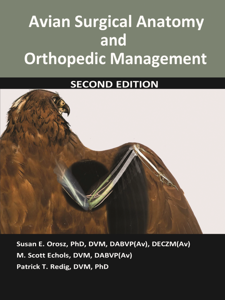
Drs Orosz, Echols and Redig worked to significantly update this book and finally publish it in 2023. While much of the original content was utilized, the new book has been substantially expanded and contains over 3 times the material. Dr Orosz updated the anatomy found on many of the beautifully illustrated plates. This information is very similar to that found in the first edition. Dr Echols invented new technology allowing researchers to see anatomical features such as blood vessels with the use of CT. The Second Edition has all new images of CT based wing and leg anatomy clearly depicting important anatomic features for several avian species. Dr Redig compiled his years of research treating avian orthopedic injuries working as the director of the Raptor Center at the University of Minnesota. Also new to the second edition are detailed descriptions of avian orthopedic injuries and their management.
The authors sincerely hope this updated book with serve those treating birds and ultimately improve the care of our feathered friends. The book can be found through numerous outlets including veterinary conferences, publishers and of course online (Amazon, etc).
Book Review: Journal of Avian Medicine and Surgery 37(4):354–356, 2023 by the Association of Avian Veterinarians
Avian Surgical Anatomy and Orthopedic Management. 2nd ed. SE Orosz, MS Echols, and PT Redig, eds. Teton NewMedia, Florence, OR, USA. 2023. 424 pp. $95.00.
There are those books that you really regret loaning out, or leaving easily accessible to sticky fingers, because they are always useful. The first edition of Avian Surgical Anatomy—Thoracic and Pelvic Limbs, by Orosz, Ensley, and Haynes was that type of book. For those that graduated veterinary school in the last century, the idea of comparative anatomy was not across classes of animals, but between orders of mammals. Yes, I found the anatomy of dogs, cats, horses, and cows fascinating because I was interested in “all creatures great and small,” but what I really craved was the opportunity to learn about the anatomy of those other classes of vertebrates: fish, amphibians, reptiles, and birds. While different texts exist for each group, the first edition of Avian Surgical Anatomy felt like it was written for a surgeon instead of an anatomist or biologist, and I enjoyed using it to prepare for my surgical cases. Once lost (or more likely stolen), I was at a loss for how to best pursue my orthopedic surgical preparations. Thankfully, those concerns have now been alleviated with the second edition of Avian Surgical Anatomy. In this updated version, not only does the title specifically note that it is for “Orthopedic Management,” but it has 2 new editors, Dr Scott Echols and Dr Patrick Redig, who are world-renowned veterinary surgeons specializing in birds. Of course, anchoring this group remains Dr Susan Orosz, whose original work captivated me with its level of detail.
The second edition is comprised of 10 chapters that cover the following topics: “Scope,” “Anatomic form and function,” “Overview of fracture management,” “General considerations for management of fractures: methods and materials,” “Anatomy of the shoulder girdle and wing,” “Management of wing fractures,” “Anatomy of the pelvic girdle and leg,” “Surgical approaches to the leg,” “Post-operative management of the orthopedic patient,” “Digital radiographs of the limbs,” and 2 appendices.
The first chapter is a single page that is meant to guide the reader to what the authors hope the reader can gain from the text. The authors reinforce to those who found the first edition to be so useful that this second edition retains many of the original illustrations used to guide us in the past, but that they are supplemented with diagnostic images, including the many computed tomography (CT) scans and arteriovenograms that Dr Scott Echols has produced and collected over the years, to reinforce how diverse these animals are and provide some granular details to guide surgeons during their approaches to fracture repair. The other important component of the scope is to introduce the additions of Dr Patrick Redig, the “godfather” of avian surgery. Dr Patrick Redig has been the leader in this field and has developed and taught veterinarians for decades. This week alone, I have used the skills I learned from reading Dr Redig’s work to coach a resident and veterinary student through surgeries on a bald eagle (Haliaeetus leucocephalus), Canada goose (Branta canadensis), and turkey vulture (Cathartes aura). The contributions of the works by both Drs Echols and Redig were quite welcomed by me and really added to what I already considered a beloved text.
In the second chapter, “Anatomic form and function,” the authors cover a range of topics, starting with a review of common anatomic terms used to describe orientation and function. This is followed by a review of important concepts regarding flight, including reviews on the feathers of the wing, wing types, aspect ratios, and concepts on aerodynamics and flight. These reviews provide the readers with insight into why it is important to correct injuries to the wing and how these injuries, or incomplete repair, may impact the recovery and flight of birds. The end of the chapter focuses on the leg and foot, again providing insight to the diversity of these structures in birds. A special section dedicated to waterfowl is also provided. This group represents a special case because they can develop severe, life-threatening injuries to their feet if not managed properly. The chapter is supplemented with 35 figures that include photographs, drawings, and diagnostic images (CT scans and radiographs) to illustrate their points. I found the images a useful supplement to the text.
Chapter 3, “Overview of fracture management,” is a short 3-page chapter that covers important concepts for the veterinary surgeon. A review of pre- and postoperative management of the orthopedic patient is provided that challenges the reader to consider the origin (wild versus captive) of the bird when assessing the type of fracture (low- versus high-impact), as well as the importance of performing a complete examination. Secondary problems, especially in wild birds, can be missed if a thorough physical examination is not performed. At my current facility, more than 40% of the raptors presenting with fractures have ocular injuries too. You never want to find out you repaired a fracture on a blind bird, and you likely will not if you pursue a thorough ophthalmologic examination on the patient at presentation. Insights into managing wounds associated with a fracture are also provided in this chapter. Two radiographic images are used to supplement the written words of this chapter.
Chapter 4, “General considerations for management of fractures: methods and materials,” is an excellent review of many of the techniques that Dr Redig has taught over the years. It includes sections on coaptation, with special commentary on the materials that should be used as well as the different methods that can be applied. Photographic images are included that cover a step-by-step approach on how to apply a body wrap and figure-of-8 bandage using different species as models. Splints are also covered, including curvededge, Altman-tape, and Shroeder-Thomas splints. The remainder of the chapter focuses on surgical fracture management. For those with limited experience in the field, the review summarizes what you will need to know from an equipment standpoint and the different procedures. A table is also provided that shows the conversion of the English to metric units for pins, a must for those with limited experience. Reviews of the primary methods used to repair avian orthopedic fractures, including intramedullary pins, external fixators, plates and nails, and the hybrid tie-in method (external
skeletal fixation and intramedullary pin) are provided. Various illustrations, including a drawing and 9 photographic images demonstrating how to tie-in the intramedullary and external skeletal pins, are used for the tie-in fixator, my preferred method. This chapter is supplemented with 14 illustrations, including drawings and photographs.
In Chapter 5, “Anatomy of the shoulder girdle and wing,” the authors provide short reviews and 138 illustrations using drawings, CT scans, and vascular contrast images of the pectoral girdle and wing. The CT and vascular scans cover a variety of species, including orange-winged Amazon parrots (Amazona amazonica), umbrella cockatoos (Cacatua alba), military macaws (Ara militaris), budgerigars Melopsittacus undulatus), pigeons (Columba livia), domestic chickens (Gallus gallus domesticus), red-tailed hawks (Buteo jamaicensis), barn owls (Tyto alba), great horned owls (Bubo virginianus), golden eagles (Aquila chrysaetos), painted storks (Mycteria leucocephala), and African geese (Anser anser domesticus). I appreciated the authors’ use of so many species across different orders to demonstrate the variability we can see in the avian wing, and this chapter should really reinforce to veterinarians the need to consider these differences when approaching their cases. The new CT images are an excellent addition to the book, and color coding is used to guide the reader about the important bones or landmarks in the images. One of the new additions I found quite useful was the inclusion of contrast images of the vascular system. While the original illustrations from the first edition are still used to demonstrate the location of blood vessels, the contrast images reveal the fine details and branching of the vessels in the wing. If you are image driven, you can spend a fair amount of time reviewing the images in this chapter and comparing them between species.
In Chapter 6, “Management of wing fractures,” the authors give step-by-step details on how to repair fractures based on the bone and location in the bone. The chapter starts by covering the fixation methods for the humerus and provides drawings and photographs to demonstrate how to perform tie-in and closed reduction repairs. The review is followed by a case that uses radiographic images to take the reader through a stepby-step approach to a tie-in. Next, the authors demonstrate how to use a tension band and tie-in repair to manage proximal and distal humeral fractures. Photographs and radiographs are again used to illustrate the authors’ points, and a second case using radiographs was used to show the reader a step-by-step approach to managing a distal humeral fracture. A similar strategy is used to guide the readers through the management of radial and ulnar fractures. Drawings are used to illustrate pin placement methods in the ulna and the techniques for managing oblique and butterfly fragments. Cases 3–14 use radiographs to demonstrate how to manage challenging radial and ulnar fractures. I appreciated these examples because there are always surgical cases that don’t follow “the book” and it is always valuable to see how other colleagues manage these cases. The remainder of the chapter focuses on metacarpal fractures and follows the same patterns as for the more proximal bones. Drawings, photographs, and radiographs are used to demonstrate how to repair the metacarpus using surgical and coaptation techniques using a series of cases (cases 15–23).
In. Chapter 7, “Anatomy of the pelvic girdle and leg,” the authors followed the same outline they used for Chapter 5, but it was for the leg. In this chapter they used 106 different illustrations to reveal the important components of the legs that a surgeon should consider when planning a procedure. Many of the same species used as models in Chapter 5 were used in this chapter too. I especially appreciated the contrast images. Being able to see the vascular patterns in the legs really gives me something to consider when approaching a repair. One of the limitations in both Chapters 5 and 7 as it relates to the CT images was that some of the legends can be challenging to read because they are imbedded in the image and have a similar grayscale.
Chapter 8, “Surgical approaches to the leg,” followed the outline of Chapter 6, where the authors start proximally and move distally through the limb. The start of the chapter focuses on the femur, and like the humeral section in Chapter 6, drawings and radiographs are used to provide the reader step-by-step approaches to repairing the bone. Additional cases are used to demonstrate how to repair distal and proximal femoral fractures. Next, the authors provide a short review on managing stifle luxation and then cases 4–10 are used to provide examples of surgical and coaptation approaches to tibiotarsal fractures. A variety of examples are used, which I think readers will greatly appreciate. The final 2 cases shared in this chapter were for managing tarsometatarsal fractures.
Chapter 9, “Post-operative management of the orthopedic patient,” provides the reader with a short review of what to expect and how to manage the avian patient postsurgery. A short introduction is followed by short passages on general considerations for the pectoral limb, immediate postoperative period (24–48 hours), passive range of motion exercises for the wing, long-term considerations, monitoring progress, postoperative issues for the pelvic limb, and beyond recovery. The chapter is short, 5 pages, but packs important information in for the reader.
The final chapter, Chapter 10, “Digital radiographs of the limbs,” uses 13 radiographic images to illustrate important landmarks on avian bones. For those with limited experience, the images will serve as a great reference for learning about these landmarks, and for those with experience, they will serve as a great quick reference.
The appendices include 2 tables, 1 that provides pin sizes for long bones in different species and a second that provides homologous terms for muscles across multiple texts. Surgeons will find Table 1 very useful because it provides intramedullary pin sizes for the humerus, radius, ulna, femur, and tibiotarsus. It also provides external skeletal fixator pin sizes and diameters for the tie-in external bar. My residents find it quite useful when preparing for surgery. A complete alphabetical index follows the appendices.
I thoroughly enjoyed reading and reviewing this text, and I was truly excited to see it in print. Because of the diversity of topics we face in our field, specialized texts, such as this one focused on surgery, are really needed to help us rise to the next level. Many of the general texts on exotic species incorporate chapters on surgery, but they are quite limited. For those with limited experience in the field, using cadavers is an excellent way to develop your surgical skills and this book provides the step-by-step approach you need to practice and gain confidence as a surgeon. For those with more experience, the text will serve as an excellent reference for surgical planning because of the diversity of species used as models. I perform orthopedic surgeries at our facility on a weekly basis but did not receive formal training in orthopedics and learned through practice, so I always find the insight of colleagues helpful to improving my skill set. The fact that this text was developed by 3 leaders in the field just increased the value of it for me.
—Reviewed by Mark A. Mitchell, Louisiana State University, School of Veterinary Medicine, Department of Veterinary Clinical Sciences, 1909 Skip Bertman Drive, Baton Rouge, LA 70803, USA
Laboratory Manual for Clinical Veterinary Technology
Oreta Marie Samples, BS, RVT, MPH, DHSc
Scott Echols, DVM, Dipl ABVP
Published 2022
Dec, 2022
‘This is a 235-page paperback, spiral bound manual for vet tech students. The manual is organized in a very logical manner and divided into clear chapters on all topics a competent vet tech should be proficient in. The manual covers domestic species (large and small) as well as some non-domestic species. Topics covered are those that apply to any species: Lab safety and zoonoses, hematology, blood smears, antigen-antibody patient side assays coagulation, coprological exam urinalysis, cytology (impression smear, fine needle aspirates, swabs) as well as large animal specific (California mastitis test and rumen fluid evaluation) and includes comments in the general sections pertinent to non-traditional species. Important procedures include necropsy, sample collection and shipping. The chapter on microbiology covers the basics that can be done in-house. Reproduction (semen analysis and vaginal cytology) is not covered.
Each chapter discusses the topic, provides figures for how to perform the procedure as well as figures to illustrate what the students are looking for and how to identify these under the microscope. The tables in each chapter are particularly useful to summarize for example the different kind of tubes for blood collection or different kinds of microbiology media: what they contain and what they are used for.
Although many clinics send out cytology to commercial laboratories, a technician who is able to properly collect and prepare and then evaluate cytology is worth a lot, because patients can be treated immediately based on preliminary assessments and is in a position to provide added value services to clients.
The manual references the AVMA CVTEA and each lab is set up to meet/ teach specific skills from the skill list. One really nice feature for the instructors is that the materials and supplies needed are listed so it is easy to prepare for the lab. An additional nice feature is that the size of the manual as well as the font are easy to read and manage while in the lab. ’
Anneke Moresco, DVM, PhD
Veterinary Technology Program Director
Western Colorado Community College
Grand Junction, CO 81505
To order, please click here