All animals possess anatomic variations that help them adapt to their environment. These variations make us all unique and beautiful.
The Grey Parrot Anatomy Project exists to discover anatomy and develop technology to better improve anatomic visualization. By understanding what is normal, we are better positioned to identify and ultimately treat the abnormal.
Discoveries made through the Project have already been used on a large variety of animals including humans. In this art series, the grey parrot, pigeon, American alligator, brown rat and human are featured. BriteVu®, a novel contrast agent developed out of the Project, was used along with computerized tomography to visualize the vasculature in each featured animal.
The heart, iris and kidney of the brown rat and upper body (American alligator) and head (all others) are shown. For more details on the project, go to www.avianstudios.com/the-grey-parrot-anatomy-project. For those interested in purchasing prints, click on the picture and it will take you to a link to the Association of Avian Veterinarians’ (AAV) website.
All proceeds are split between the AAV Research Fund and the Grey Parrot Anatomy Project Research Fund, both of which are tax deductible donations.
M. Scott Echols, DVM, Dipl ABVP (Avian Practice)
Project Director
Grey Parrot Anatomy Project
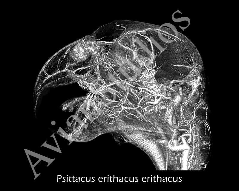
Grey Parrot (Psittacus erithacus erithacus)
Grey Parrot (Psittacus erithacus erithacus)
The Grey Parrot Anatomy Project has been established in part to improve upon and create technology that allows the world to study anatomy of any animal. While the focus is on the grey parrot, the methodology being developed has already been used on birds, mammals (including humans), reptiles and more. One of the advances has been the development of BriteVu®, a new contrast agent that allows researchers to see vasculature (with the aid of a CT scan) down to the capillary level. A grey parrot head is seen here with vessels highlighted using BriteVu®. One interesting finding has been the tortuous vessels at the base of the head. These vessels have slack to allow the bird to turn its head around without significantly altering blood flow.
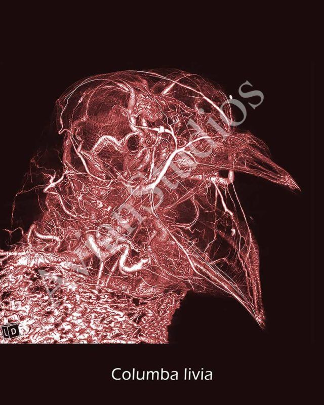
Pigeon (Columba livia)
Pigeon (Columba livia)
The Grey Parrot Anatomy Project has been established in part to improve upon and create technology that allows the world to study anatomy of any animal. While the focus is on the grey parrot, the methodology being developed has already been used on birds, mammals (including humans), reptiles and more. One of the advances has been the development of BriteVu®, a new contrast agent that allows researchers to see vasculature (with the aid of a CT scan) down to the capillary level. In this pigeon perfused with BriteVu®, the collar plexus of the neck subcutaneous veins is just visible at the bottom of the picture. This vascular plexus helps the pigeon thermoregulate by having an extensive blood supply close to the skin.
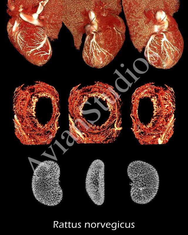
Brown Rat (Rattus norvegicus)
Brown Rat (Rattus norvegicus)
The Grey Parrot Anatomy Project has been established in part to improve upon and create technology that allows the world to study anatomy of any animal. While the focus is on the grey parrot, the methodology being developed has already been used on birds, mammals (including humans), reptiles and more. One of the advances has been the development of BriteVu®, a new contrast agent that allows researchers to see vasculature (with the aid of a CT scan) down to the capillary level. In this image, the rat heart (top), iris (middle) and kidney (bottom) are perfused with BriteVu® revealing the microvasculature of each organ. By seeing the vasculature, researchers can study the effect of cancer, diabetes mellitus, infections, autoimmune disorders and much more on the blood vessels.
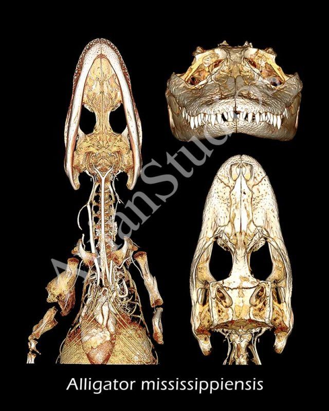
American Alligator (Alligator mississippiensis)
American Alligator (Alligator mississippiensis)
The Grey Parrot Anatomy Project has been established in part to improve upon and create technology that allows the world to study anatomy of any animal. While the focus is on the grey parrot, the methodology being developed has already been used on birds, mammals (including humans), reptiles and more. One of the advances has been the development of BriteVu®, a new contrast agent that allows researchers to see vasculature (with the aid of a CT scan) down to the capillary level. The vasculature of the alligator is highlighted using BriteVu® with the heart and its major vessels visualized in left image. The alligator also has osteomyelitis of the right humerus.
Human (Homo sapiens)
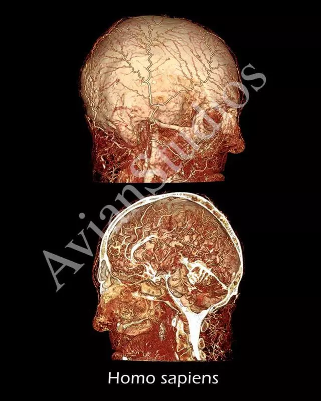
Human (Homo sapiens)
The Grey Parrot Anatomy Project has been established in part to improve upon and create technology that allows the world to study anatomy of any animal. While the focus is on the grey parrot, the methodology being developed has already been used on birds, mammals (including humans), reptiles and more. One of the advances has been the development of BriteVu®, a new contrast agent that allows researchers to see vasculature (with the aid of a CT scan) down to the capillary level. Dr Bruce Wainman of McMaster University in Canada worked with the body donor program to perfuse this human head with BriteVu®. This was the first CT data set that demonstrated the blood vessels of the human head with such great detail. Images such as these are re-writing our understanding of anatomy even in those species that have been regularly studied for over a thousand years.
Northern Helmeted Curassow (Pauxi pauxi)
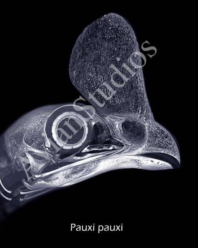
Northern Helmeted Curassow (Pauxi pauxi)
The Grey Parrot Anatomy Project has been established in part to improve upon and create technology that allows the world to study anatomy of any animal. While the focus is on the grey parrot, the methodology being developed has already been used on birds, mammals (including humans), reptiles and more. One of the advances has been the development CT (CAT scan) protocols for use in many species. Animals, such as this northern helmeted curassow, are scanned in and the data is then stored for use by researchers worldwide. The inside spongy bone of the curassow’s casque, situated on top of the upper beak, is readily visualized using this micro CT protocol.